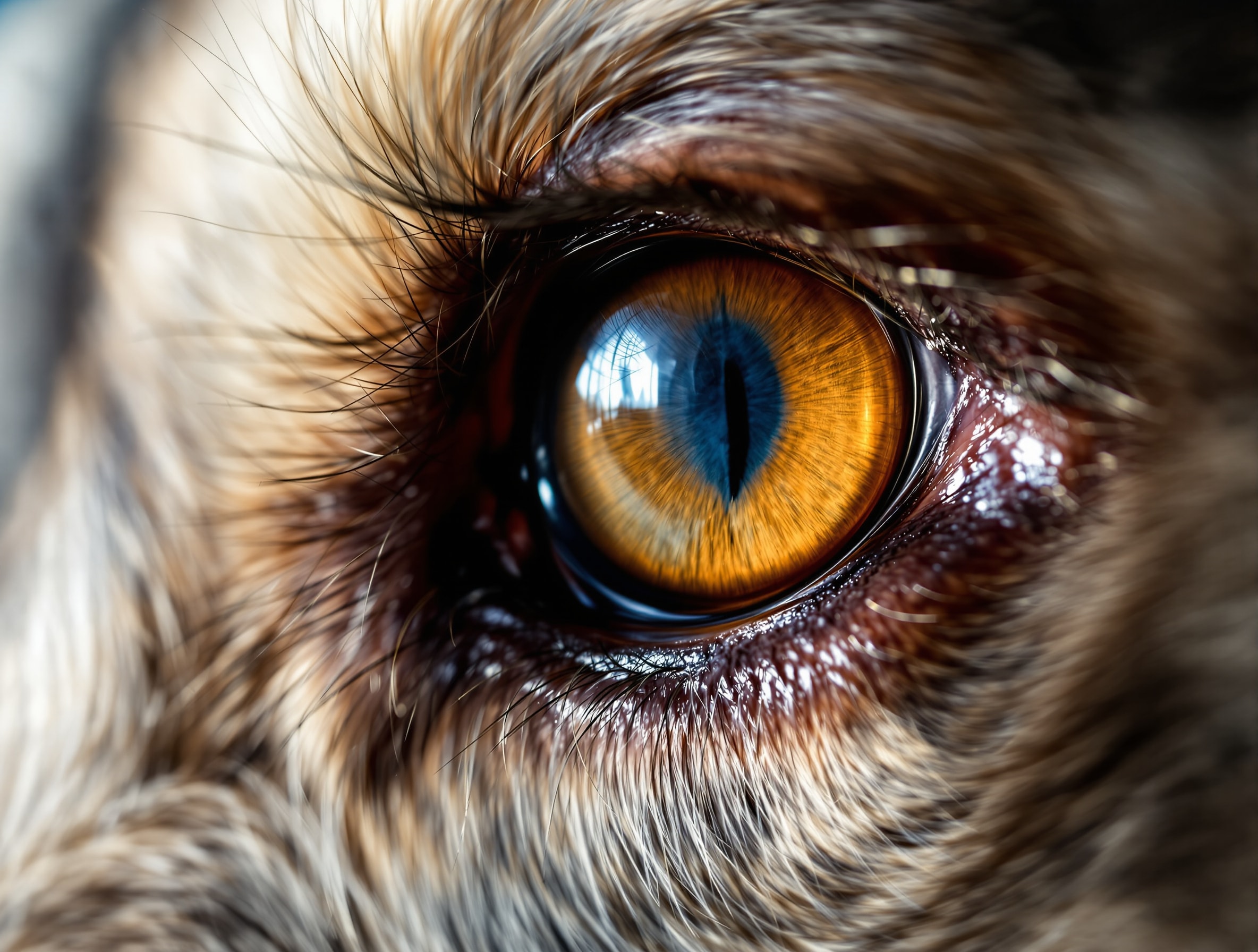
Pannus and the Greyhound
Pannus, or Chronic Superficial Keratitis, is an immune mediated disease of the eyes with which Ray and Porcia were both afflicted. It can cause blindness if it is not effectively treated. Pannus is not as common a disease in Greyhounds as it is in some other breeds, which is part of the problem: it is easy for it to go unrecognized for a while in a Grey.
We are not experts in veterinary medicine. We are two people who lived with two Pannus dogs for years, learned the routine the slow way, and kept this page up in the hope that some of it is useful to somebody else in the same position.
The One Thing to Take Away From This Page
If you take away one piece of knowledge from this page it should be that Pannus requires specialized treatment. The disease can be controlled in the great majority of cases, but even excellent general veterinarians simply do not see enough of it to manage it well. Regular visits to a veterinary ophthalmologist must be made to be sure a Pannus dog is receiving the proper levels of the appropriate medications.
The reason this matters so much is that the margin is narrow at both ends. Too little medication and the disease advances across the cornea. Too much, applied for years on end, and long-term corticosteroid use in the eye can bring on corneal dystrophy, so that the treatment for the first disease has quietly created a second one. Getting that dose right, and adjusting it as the disease and the seasons change, is a specialist's job. The American College of Veterinary Ophthalmologists is where to start looking for one.
Ray's Story
Ray languished in a Florida Greyhound rescue kennel for several months before we adopted him. We believe he was not adopted sooner because of his Pannus, since there were plenty of healthy dogs available and he was nearly five years old besides. When we adopted him the disease was very active in both eyes and he was in real peril of losing his sight.
We went into it apprehensive and largely ignorant, which we would not recommend as a method. What saved him was not us. It was getting him in front of a veterinary ophthalmologist quickly and then never letting the schedule slide. In time his condition came under control and his eyesight was saved, and he went on to spend the rest of his life as a working therapy dog, which is not a career available to a blind dog.
What Pannus Is
Pannus is a slowly progressive disorder of the canine cornea, characterized by pigmentation, vascularization and opacification. In plain terms, the clear window at the front of the eye is gradually invaded: first by inflammatory cells, then by new blood vessels growing into tissue that is normally free of them, then by scar tissue, and finally by brown pigment. Each of those layers makes the cornea a little less transparent. Left alone long enough, the window closes.
It is seen predominantly in one large herding breed, with sporadic cases in other breeds and in mixed breeds as well. Greyhounds are not a high-incidence breed, so a Greyhound with Pannus is unusual enough that it can be missed or mistaken for something more ordinary in a general practice.
What Causes It
The cause is not known, and several factors are probably involved at once:
- Heredity. The strong breed incidence suggests a heritable predisposition.
- Ultraviolet radiation. UV exposure appears to be an important inciting and propagating factor. Dogs living at high altitude and low latitude are consistently more severely affected, and this is the one factor an owner can actually do something about.
- Immune response. The disease is immune mediated. The reaction may be directed against foreign material at the surface of the eye, or against the corneal tissue itself.
- Infection. A long list of infectious agents has been blamed over the years, and none of them has held up as a significant cause.
Worth saying plainly: it is nobody's fault, it is not contagious, and it is not caused by anything you did or failed to do.
What It Looks Like
The first sign is usually a haze creeping in from the edge of the clear cornea, white, pink or brownish, most often starting at the outer lower corner of the eye and extending inward. Before that there may be redness and brown pigment in the membrane just outside the cornea. As it advances, the whitish infiltrate is followed by visible fine blood vessels growing in across the surface, then by a duller, thickened, tissue-like appearance, and eventually by brown pigment.
A smaller number of dogs show one or both of two additional signs: a thickened, reddened, depigmented third eyelid, or chronic shallow ulceration of the lower lid near the inner corner. An eye that is actively progressing may be uncomfortable. An eye that is well controlled generally is not, which is a useful thing to know, because a Pannus dog on treatment who suddenly starts squinting, holding an eye shut or pawing at it is telling you something has gone wrong. That is a same-day call to the vet, not a wait-and-see.
How It Is Diagnosed
Diagnosis is clinical. A veterinary ophthalmologist examines the eye under magnification, looks at the pattern and location of the changes, stains the cornea to rule out ulceration, and checks tear production, because dry eye can look superficially similar and is treated quite differently. In a high-incidence breed the picture is typical enough that the diagnosis is straightforward. In a Greyhound it is worth the specialist appointment to be certain what you are treating. The veterinary manual's overview of corneal disorders in dogs is a good place to see how many quite different problems present in a similar way.
How It Is Treated
There is no cure, and this is the part that takes new owners a while to absorb. What there is, in the vast majority of cases, is good control: the progress of the disease can be halted and the eye kept stable, especially if treatment starts early. The early changes, the cell infiltrate and the blood vessels, usually reverse with treatment. The late changes, scar tissue and pigment, tend to be permanent once they have arrived. That asymmetry is the whole argument for catching it early.
Treatment rests on two things:
- Topical immune suppression, for life. Anti-inflammatory eye drops or ointment, typically a topical immunomodulator and a corticosteroid in some combination, applied on a fixed daily schedule and never stopped. Even a short interruption of a few weeks can produce a severe relapse that costs vision permanently. Doses are adjusted by the ophthalmologist over time, and are often stepped up in summer and eased back in winter.
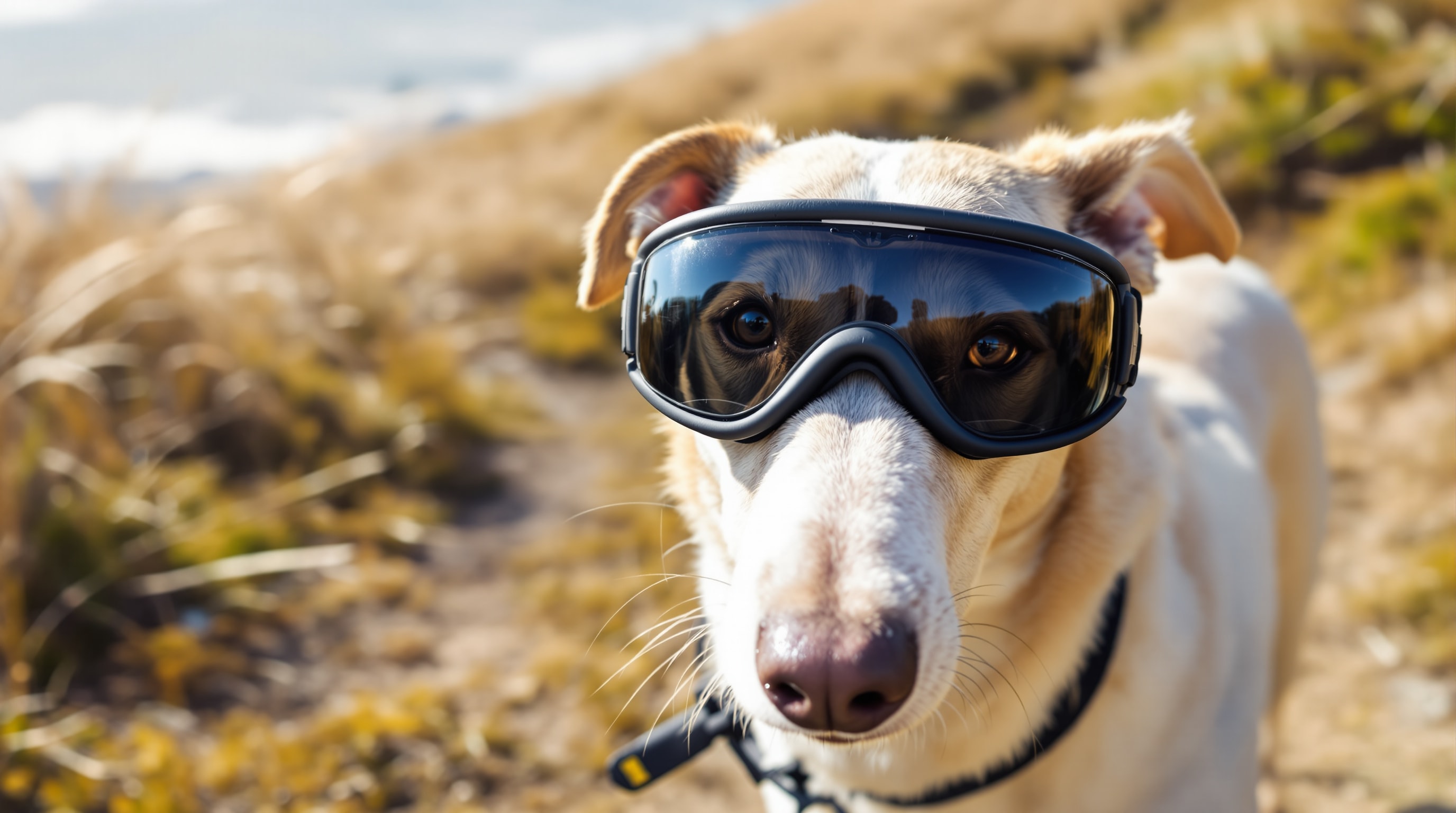
- Protection from ultraviolet light. Since UV drives the disease, reducing exposure genuinely reduces the medication needed. That means shade, avoiding the brightest hours in summer, thinking hard about high altitude trips, and in some cases protective dog goggles. Yes, they look ridiculous. Yes, they work, and a surprising number of dogs accept them within a week if you introduce them gently.
Other measures exist for cases that resist the standard approach, including beta-irradiation and surgical removal of the affected superficial corneal layer, but these are unusual, not widely available, and firmly a specialist's decision rather than an owner's.
Reexaminations should be followed closely. Early on they may need to be every couple of months; once medical control is established they can often be stretched out considerably. Between scheduled visits, any change in the appearance of the eye is a reason to call.
Living With a Pannus Dog
The clinical description makes it sound harder than it is. Day to day, what Pannus actually means is a small ritual repeated twice a day forever. Some things we learned:
- Attach the drops to something you already do without fail, morning and evening. Missed doses are the main way well-controlled dogs lose ground, and they are almost always the result of an unstructured routine rather than a forgetful owner.
- Medicate before the meal, not after. The dog will forgive almost anything that is immediately followed by dinner.
- Keep a spare bottle. Running out on a holiday weekend is the classic way to open a two-week gap, and a two-week gap is exactly what you have spent years avoiding.
- Take a photograph of each eye in the same light every month or so. Gradual change is invisible when you look at it every single day, and a folder of pictures makes it obvious.
- Keep every ophthalmology report in one place and take them all to each visit, particularly if you ever move or change practices.
- Budget for it. Specialist ophthalmology visits and lifelong topical medication are a real recurring cost, and it is far better to know that going in than to discover it in year two.
- Watch for pain. Squinting, an eye held shut, pawing or a sudden change in appearance means stop and call, especially for a dog on long-term steroid drops.
None of this stopped either of our dogs doing anything. Ray worked as a therapy dog for years with two treated eyes, and Porcia's Pannus was, in the accounting of her long and comfortable retirement, a minor line item.
What This Page Is and Is Not
This is one family's experience of one disease in two dogs. It is not veterinary advice, it is not a diagnosis, and nothing here should be used as a reason to delay or to alter what your own veterinarian has told you. If you suspect your dog has Pannus, or if you have been given the diagnosis by a general practice and have not yet seen a specialist, please see a veterinary ophthalmologist. That single step is the one that made the difference here.
Ray's own page tells the rest of his story, and there is more about the breed generally on our Greyhound facts page.